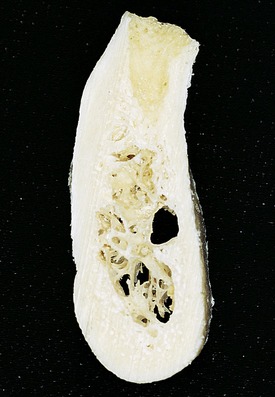
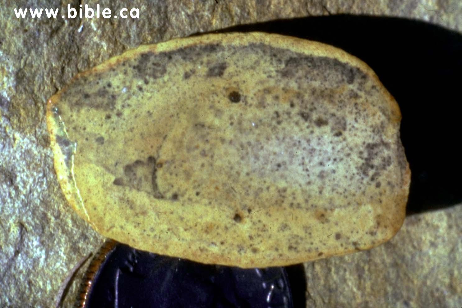
Cross Section Of A Bone / Fossilized Human Finger: Found in a formation famous for ... : Internal structure of a human long bone, with a magnified cross section of the interior.. Related posts of cross section of human bone diagram muscles and bones of the human body. While it is not as hard as compact bone, spongy bone plays an important role of protecting the marrow where blood cells are produced. This is known as the periosteum. Cross section of long bone. 100x first focus in the compact decalcified bone (cb) on the left part of the image, you can see small dots, which are.
It consists of two layers; Compact bone, also called cortical bone, dense bone in which the bony matrix is solidly filled with organic ground substance and inorganic salts, leaving only tiny spaces (lacunae) that contain the osteocytes, or bone cells.compact bone makes up 80 percent of the human skeleton; Cartilaginous area at the ends of long bones where lengthwise growth takes place in the immature skeleton. End of a long bone. Marrow in the shaft of long bones is typically yellow, with red marrow in the head through the cancellous bone.
14: INFERIOR ALVEOLAR NERVE LATERALIZATION AND MENTAL ... from pocketdentistry.com Each bone in your body is made up of three main types of bone material: Related posts of cross section of human bone diagram muscles and bones of the human body. There are three general classes of bone. 100x first focus in the compact decalcified bone (cb) on the left part of the image, you can see small dots, which are. This slide contained a cross section of a very small bone, and you are looking at the entire thickness of the shaft of the bone. After a fracture, woven bone forms initially and is gradually replaced by lamellar bone during a process known as bony substitution. They are obtained by taking imaginary slices perpendicular to the main axis of organs, vessels, nerves, bones, soft tissue, or even the entire human body. Cross‐sectional area is derived from the integral of the bone mass profile across the narrow region.
Bone cross section view :
Muscles and bones of the human body 12 photos of the muscles and bones of the human body anatomy bones of the human body quiz, major muscles and bones in the human body, muscles and bones in the human body, number of muscles and bones in the human body. The periosteum, or outside skin of the bone; And why does the marrow stop where it does, and so sharply? Browse 53 bone marrow cross section stock photos and images available, or search for bone cross section or bone cells to find more great stock photos and pictures. The remainder is cancellous bone, which has a spongelike appearance with numerous large spaces and is found in the. Cross section of mandible at first molar region showing cortical and spongy bone basic concepts in osteogenesis bone is a dynamic biological tissue, composed of various metabolically active cells that are integrated into a rigid framework. Studies on the cross‐sectional geometry of long bones in african apes have documented that shape ratios derived from second moments of area about principle axes (e.g., i max /i min) are often correlated with habitual locomotor behaviors.for example, humeral cross‐sections tend to appear more circular in more arboreal and forelimb suspensory chimpanzees compared with terrestrial quadrupedal. The upper (biting) surfaces of the tooth are at top, with the lower sections (bottom) embedded in the gums and jaw bone (not shown). Learn vocabulary, terms and more with flashcards, games and other study tools. This slide contained a cross section of a very small bone, and you are looking at the entire thickness of the shaft of the bone. 100x first focus in the compact decalcified bone (cb) on the left part of the image, you can see small dots, which are. The large dark spots are passages for blood vessels and nerves. The compact bone is made up of osteon.
Terms in this set (3) epiphysis. The cortical bone equivalent area of the cross‐section of the region of interest (femoral neck or shaft), with all soft tissue voids (trabecular and cellular spaces) eliminated (cm 2). Cartilaginous area at the ends of long bones where lengthwise growth takes place in the immature skeleton. Compact bone, spongy bone, and bone marrow. Related posts of cross section of a long bone.
"Bone Cross Section" for Radius Digital Science on Behance from mir-s3-cdn-cf.behance.net Foot bone anatomy x ray 12 photos of the foot bone anatomy x ray foot bone anatomy x ray, bone, foot bone anatomy x ray. Learn vocabulary, terms and more with flashcards, games and other study tools. Marrow in the shaft of long bones is typically yellow, with red marrow in the head through the cancellous bone. Related posts of cross section of a long bone. To the left is muscle tissue, and to the right is bone marrow. It consists of two layers; Bones of the human head Cartilaginous area at the ends of long bones where lengthwise growth takes place in the immature skeleton.
Human bone, cross section diagram of femur showing osteon, veins, marrow.
Terms in this set (3) epiphysis. Compact bone, also called cortical bone, dense bone in which the bony matrix is solidly filled with organic ground substance and inorganic salts, leaving only tiny spaces (lacunae) that contain the osteocytes, or bone cells.compact bone makes up 80 percent of the human skeleton; Learn vocabulary, terms and more with flashcards, games and other study tools. If we were to cut the femur bone in half, we would see that it contains various layers. Cross section of long bone. The compact bone is made up of osteon. Table 1 describes the bone markings, which are illustrated in (figure 4). 100x first focus in the compact decalcified bone (cb) on the left part of the image, you can see small dots, which are. This slide contained a cross section of a very small bone, and you are looking at the entire thickness of the shaft of the bone. The surface features of bones vary considerably, depending on the function and location in the body. There are three general classes of bone. Internal structure of a human long bone, with a magnified cross section of the interior. Browse 53 bone marrow cross section stock photos and images available, or search for bone cross section or bone cells to find more great stock photos and pictures.
Foot bone anatomy x ray 12 photos of the foot bone anatomy x ray foot bone anatomy x ray, bone, foot bone anatomy x ray. After a fracture, woven bone forms initially and is gradually replaced by lamellar bone during a process known as bony substitution. There are three general classes of bone. There are trabeculae in spongy bone which gives its sponge like appearance. Related posts of cross section of a long bone.
Fossilized Human Finger: Found in a formation famous for ... from www.bible.ca Internal structure of a human long bone, with a magnified cross section of the interior. Related posts of cross section of a long bone foot bone anatomy x ray. I don't find it enhances the image. Start studying cross section of bone. Why is the marrow red? Two types of bone tissues in cross section of a long bone : It consists of two layers; 100x first focus in the compact decalcified bone (cb) on the left part of the image, you can see small dots, which are.
They are obtained by taking imaginary slices perpendicular to the main axis of organs, vessels, nerves, bones, soft tissue, or even the entire human body.
As the names suggest compact bone looks compact and the spongy bone looks like sponges. They are obtained by taking imaginary slices perpendicular to the main axis of organs, vessels, nerves, bones, soft tissue, or even the entire human body. It consists of two layers; To the left is muscle tissue, and to the right is bone marrow. There are three general classes of bone. Table 1 describes the bone markings, which are illustrated in (figure 4). Learn vocabulary, terms, and more with flashcards, games, and other study tools. Bones of the human head They are obtained by taking imaginary slices perpendicular to the main axis of organs, vessels, nerves, bones, soft tissue, or even the entire human body. After a fracture, woven bone forms initially and is gradually replaced by lamellar bone during a process known as bony substitution. The large dark spots are passages for blood vessels and nerves. The surface features of bones vary considerably, depending on the function and location in the body. Related posts of cross section of human bone diagram muscles and bones of the human body.